
Eight models across three scales. Peel the parts off one at a time, cut it open on any plane, put the crowding back in, fly through the cytosol, run it through its own division — then take the oxygen away and watch, in order, exactly what fails.
Android. Closed testing now, Google Play soon.
Everything in the app is a surface, and almost every surface is the same surface. The plasma membrane, the nuclear envelope, the ER, every Golgi cisterna, every vesicle and lysosome and both mitochondrial membranes are one material — a lipid bilayer five nanometres thick — folded into different shapes. A cell is not a bag of parts. It is one kind of film wrapped an enormous number of times, and the parts are what you call the pockets it makes.
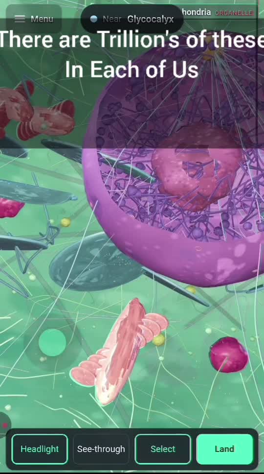
Cut a cell and you are looking into a bag, not at a sawn solid. The cut is a clipping plane and the faces are deliberately not capped, because there is nothing to cap. What you see is what a real section shows.
The cytosol is 200–300 mg/mL of protein. Every diagram ever printed shows a nearly empty bag. One molecule in about six thousand is drawn here, at true size, and the note says exactly that.
Lit, fluorescence and electron. These genuinely disagree about what is there, and switching between them on one cell is the lesson.
Chromatin condensing, the envelope falling into the ER and being reissued, the spindle built from the same tubulin the interphase array came down from. The slider is not linear in time — 23 of 24 hours are interphase — and the readout carries the real elapsed time so the distortion stays visible.
Secretion, endocytosis, gene expression, respiration, and the proton circuit inside a mitochondrion — as routes with something moving along them.
One world unit is 1 µm. The scene spans four orders of magnitude, from a 90 µm neuron to a 40 nm patch of membrane.
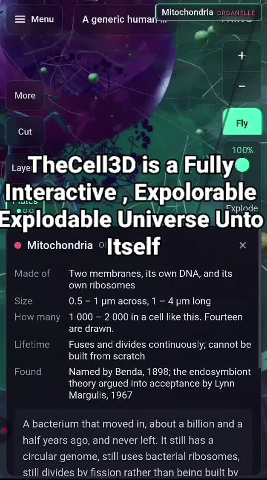
Each model carries its structures, its real numbers, camera bookmarks and a scripted tour. Every structure's name, colour, opacity and note is editable, and stays that way.
| Model | Scale | Why it is here |
|---|---|---|
| Somatic cell | 20 µm | The parts nearly every human cell shares |
| Neuron | 90 µm | Everything built in the last 20 µm, and carried |
| Red blood cell | 8 µm | Threw away its nucleus; can't use the oxygen it carries |
| Heart muscle cell | 46 µm | Up to 40% mitochondria, and no way to stop for breath |
| Nucleus | 6 µm | Two metres of DNA, filed, behind three thousand doors |
| Mitochondrion | 2 µm | A bacterium that moved in, selling surface area |
| Membrane patch | 40 nm | The material everything else is folded out of |
| E. coli | 2 µm | No rooms at all, and winning for 3.5 billion years |
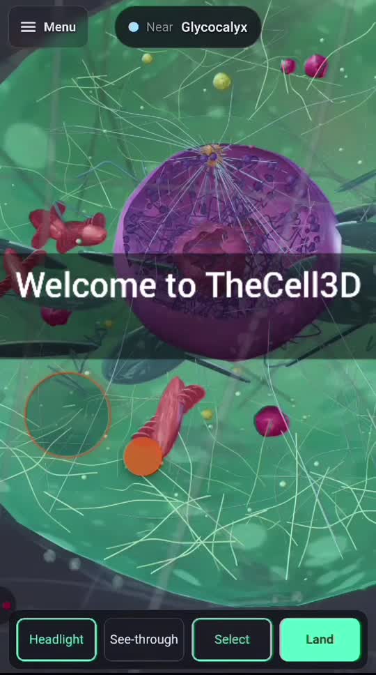
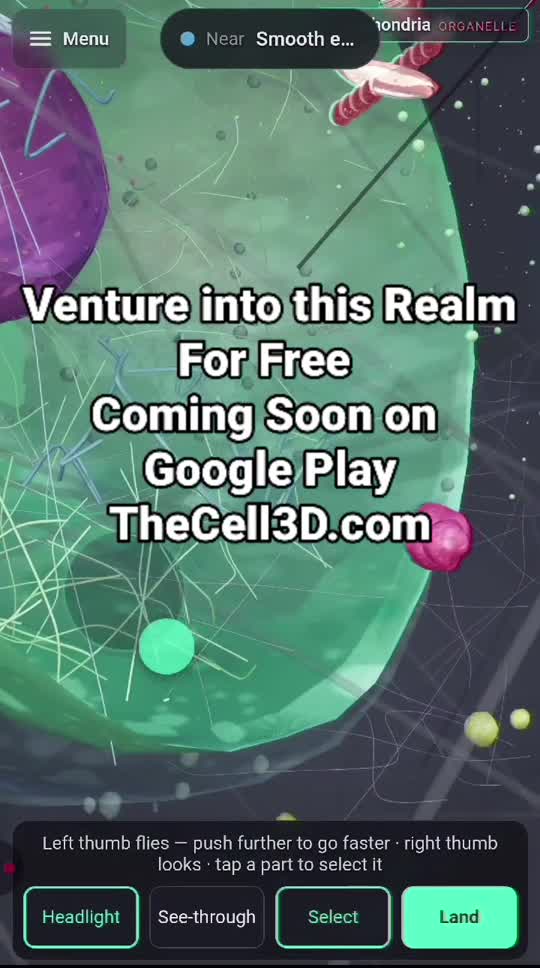
Left thumb flies, right thumb looks, tap a part to select it. Every piece a structure is made of is merged into one geometry, so the crowded cytosol costs what a single mesh costs.
Failure Understanding Simulation Engine. The same engine that runs component failure in CircuiTry3D, given a cell to work on. The domain is the ischaemic cascade, where each organelle's failure is the next one's environment.
A damaged cell dies tidily and is eaten with no inflammation. A cell whose ATP has run out cannot do that — it swells, bursts and inflames the tissue. Same insult; the difference is whether there was enough energy left to die properly.
Damaged DNA with p53 intact gives arrest, then apoptosis. The same damage with p53 lost gives a cell that divides anyway. Turn the dial down and F.U.S.E. stops calling it a death and starts calling it a lineage.
TheCell3D is in closed testing on Google Play. Testers get the full app, free, and everything they report goes into the build. Teachers especially — if it doesn't survive a classroom it isn't finished.
Send the Google account email you use on your Android phone. That address is what Play needs to let you in.